
When doctors need to see what’s happening inside your body, they rarely rely on just one type of scan.
Multi-modality imaging combines different scanning techniques to create a more complete diagnostic picture.
This approach is like using multiple camera lenses to photograph a landscape – each captures different details that together form a comprehensive view.
By integrating various imaging methods, healthcare providers can see problems that might be missed with a single scan, leading to more accurate diagnoses and better treatment plans for you.
What Makes Multi-Modal Imaging Special?
Multi-modal imaging brings together different scanning technologies that each have unique strengths. When combined, they overcome individual limitations and provide complementary information.
Think of it like investigating a crime scene – one detective might look for fingerprints while another analyzes footprints. Together, they build a more complete case than either could alone.
The integration of various scanning techniques offers several key benefits:
- Enhanced accuracy in detecting and characterizing abnormalities
- Reduced false positives and negatives that can occur with single-modality approaches
- More comprehensive understanding of anatomical and functional relationships
- Better planning for treatments like surgery or radiation therapy
Common Imaging Modalities and Their Complementary Roles
Different scanning techniques reveal different aspects of your body’s structures and functions. Here’s how some of the most common ones work together:
| Imaging Technique | What It Shows Best | Limitations | Complementary Partner |
| CT Scan | Bone structure, dense tissues | Limited soft tissue contrast | MRI for soft tissue details |
| MRI | Soft tissues, neural pathways | Time-consuming, less detail on bones | CT for bone structure |
| PET | Metabolic activity, cellular function | Lower anatomical resolution | CT/MRI for anatomical context |
| Ultrasound | Real-time movement, blood flow | Limited depth penetration | CT/MRI for deeper structures |
When doctors use these techniques together, you get the benefit of each method’s strengths while minimizing their individual weaknesses.
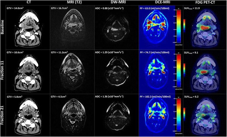
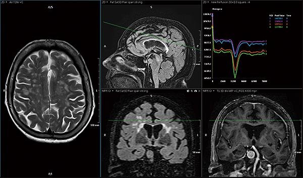
How Fusion Imaging Works in Practice?
Modern diagnostic centers now routinely combine different imaging methods to create fused images that overlay information from multiple scans.
For example, PET-CT fusion overlays metabolic information from PET scans onto the detailed anatomical framework provided by CT images.
This helps your doctor pinpoint exactly where abnormal cell activity is occurring in relation to surrounding structures.
Similarly, MRI-ultrasound fusion is revolutionizing prostate cancer detection.
The real-time guidance of ultrasound is combined with the detailed tissue characterization of MRI, allowing for more precise biopsies that are twice as likely to detect significant cancers compared to conventional methods.
Looking to the Future of Diagnostic Imaging
The field continues to evolve rapidly. Artificial intelligence is now being used to analyze multi-modal images, finding patterns and correlations that might be missed by the human eye alone.
Future developments in integrated imaging include:
New contrast agents that work across multiple imaging platforms Faster scanning times that reduce your time in the machine Smaller, more portable devices that bring advanced scanning to remote locations Reduced radiation exposure while maintaining or improving image quality.
Making the Most of Your Diagnostic Imaging Experience
When your doctor recommends imaging tests, you can ask if a multi-modal approach might be beneficial for your specific condition.
Understanding that different scans serve different purposes can help you appreciate why multiple tests might be necessary.
Remember that integrated imaging isn’t just about getting more tests – it’s about getting the right combination of tests to create the most comprehensive diagnostic picture for your unique situation.
The Complete Picture
Multimodal imaging represents one of the most significant advances in modern diagnostic medicine.
By combining various scanning techniques, doctors can now see your body’s structures and functions with unprecedented clarity and detail.
This integrated approach means more accurate diagnoses, better treatment planning, and improved outcomes for you as a patient.
As technology continues to advance, the seamless integration of multiple imaging methods will become even more sophisticated, further enhancing our ability to see beyond the surface and understand the complex workings of the human body.